
|
To understand what a crown lengthening is, it is important to understand the structure of a tooth. The crown is the visible part of our teeth in the mouth. The neck of the tooth is usually surrounded by our gum and the root of the tooth is secured in the jaw bone. |

With a crown lengthening procedure, the gum is gently lifted away from the defect site to gain access to the bone structure surrounding the tooth. Some of the bone structure is then removed around the neck of the tooth to enlarge the exposed area of the tooth. Excess gum is trimmed away, and the remaining gum is resecured around the tooth and left to heal. Once the sutures are removed and the area is healed, restorations can then be done on the new, extended tooth structure.


|
A Gingivectomy is the total removal of the gum/gingiva in and/or around teeth to treat gum disease or to increase the height and/or width of teeth. Some patients are born with excessive gum growth and others present with it due to trauma, hormonal changes, medications, orthodontic work, and other factors. |

|
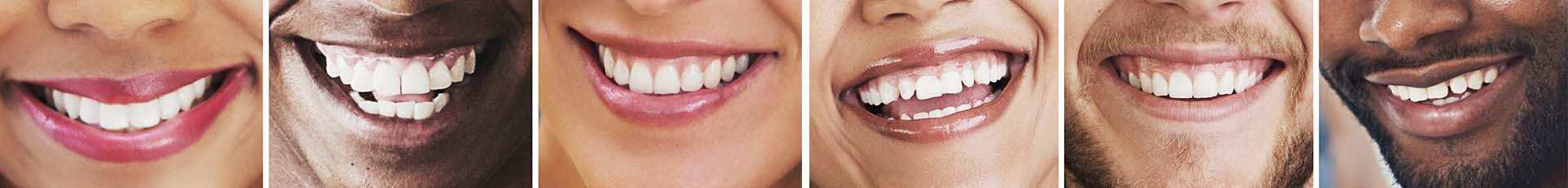
A Gingivaplasty is done to shape the gum in specific areas which present with an uneven gum line to make the gum appear more natural. Although this is usually done for patients who simply want their gums to look better, there are instances where it is a requirement for the patient’s better health. |

|
A Soft Tissue Graft is usually done when a patient presents with bone and soft tissue loss (recession) which makes the teeth look longer and is generally not aesthetically pleasing to look at. This could be on one or more teeth. There are many factors which could cause gum recession such as trauma and previous orthodontic work, however periodontal disease remains the most common factor. With periodontal disease, the bacteria eat away at the bone surrounding our teeth and the longer it is left untreated, the more severe the defects will become. Bone keeps our gum around the teeth, thus when bone loss occurs, our gum recedes (pulls away) to where the bone level is. If left untreated, the condition will worsen over time, and could lead to tooth loss. |
By performing a soft tissue graft(s) on recessed teeth, we can restore lost bone and gum. If the recession is caused by gum disease, the first step will be to bring the infection under control and to maintain a stable environment for at least 3 months before surgery can be performed.
Once a healthy foundation is created, soft tissue can be harvested – usually from the palate – and grafted/transplanted into the defect site. Bone regeneration can also be done to regenerate new bone growth in the sites for a better long-term prognosis.

|
A frenum is a little piece of soft tissue that connects our gum and lip line, most easily seen between the front lower and top teeth. We also have several other frenums in our mouths which can be seen when pulling the lips away from the gum, or when lifting the tongue. |
When a frenum is too big, causing obstruction of movement, gum recession or teeth to drift apart (diastema) it is referred to as an active frenum and it can be treated with a frenectomy procedure. An active frenum under the tongue can hinder speech and eating, and also causes a problem for suckling babies as they cannot use their tongues adequately to latch onto the feeding mother. Active frenums between the teeth can cause gingival recession, diastemas and hinder orthodontic work, thus patients are often referred for a frenectomy before orthodontic work may commence.
Although there are rare extreme cases which require surgical severing of the frenum attachment, in most cases the frenectomy can be done with a laser. By using a laser, the procedure is quick and painless, and the wound does not need sutures to be placed because the laser immediately seals off the wound. Post-operative discomfort is little to none and the wound heals extremely fast.
A frenectomy can be done in the dental chair and takes around 20 – 30 minutes.